
🐶 Legg Calve Perthes Disease in Dogs
Lateef Bhatti
Author
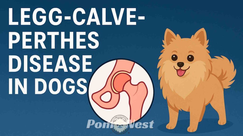
Legg Calve Perthes Disease in Dogs is a degenerative orthopedic condition, common in young, small breed dogs, caused by the interruption of blood supply to the femoral head (ball of the hip joint). This lack of blood flow leads to bone death (avascular necrosis), resulting in the collapse of the femoral head, severe pain, and progressive lameness. Treatment for this painful condition almost always involves surgery, typically a femoral head ostectomy (FHO).
What Is Legg Calve Perthes Disease in Dogs?
If your dog is limping or showing signs of hip pain, you might worry about major conditions like hip dysplasia. However, another serious orthopedic dog condition, Legg-Calve-Perthes disease (LCPD), is common in smaller breeds and warrants immediate veterinary attention. This disease affects the hip joint, specifically the top of the thigh bone, which is called the femoral head. When a dog develops Legg Calve Perthes disease in dogs, the bone cells die off, which leads to joint collapse and severe pain.
Table of Contents
ToggleThe Mechanics of LCPD: What Happens to the Hip Joint?
The hip is a classic ball-and-socket joint, where the femoral head (the ball) sits neatly inside the acetabulum (the socket). In LCPD, the blood supply to the femoral head temporarily or permanently stops. This lack of blood flow causes the bone tissue to undergo a process called femoral head necrosis (bone cell death). As the bone dies, it weakens, cracks, and eventually collapses, causing the ball to flatten and disintegrate. This results in significant friction, inflammation, and rapid hip joint degeneration, eventually leading to painful osteoarthritis in dogs.
Which Breeds Are Most Susceptible?
LCPD is primarily a disease of small-breed dogs, although it can occasionally affect larger dogs. Breeds that are most commonly diagnosed include Yorkshire Terriers, West Highland White Terriers, Miniature Poodles, and Cairn Terriers. If you own a small-breed puppy, it’s important to monitor their development and gait closely for any early signs of canine lameness.
The Typical Age of Onset
The signs of LCPD usually appear during the puppy stage, before the dog reaches skeletal maturity. Most dogs show symptoms between five and eight months of age. Because the hip joint is still rapidly developing during this period, the disruption to the bone’s blood supply causes catastrophic failure.
Causes of Legg Calve Perthes Disease in Dogs

Understanding the underlying cause of LCPD is critical for both diagnosis and prevention. Researchers don’t know the exact trigger, but they believe a combination of factors is responsible for initiating this painful condition.
Genetic Factors and Hereditary Links
The most significant cause appears to be genetics. LCPD is strongly suspected to be an inherited condition, meaning dogs carrying the specific genes are predisposed to developing it. This link is why certain small breeds suffer from the condition much more frequently than others. Breeders of at-risk breeds should screen their lines carefully to prevent passing the trait down.
The Critical Role of Blood Supply (Avascular Necrosis)
The immediate mechanism that destroys the femoral head is the interruption of blood flow, known as avascular necrosis. Without a continuous supply of oxygen and nutrients carried by the blood, the bone cells simply cannot survive. The cause of this blood flow interruption remains a topic of scientific debate, but researchers suspect either a structural defect in the blood vessels or a blockage causes the issue.
Environmental or Nutritional Influences
While genetics are key, environmental factors may play a smaller supporting role. For example, some early theories suggested trauma or hormonal imbalances might trigger the onset. However, most experts now agree that while trauma to the hip might worsen an already deteriorating joint, it likely doesn’t cause the underlying Legg-Calve-Perthes disease. Nutritional issues are generally discounted as a primary cause, but maintaining a balanced diet is always important for healthy joint development.
Symptoms of Legg Calve Perthes Disease in Dogs

The symptoms of LCPD are usually progressive, meaning they start subtly and worsen over a few weeks or months as the hip joint collapses further.
Early Signs: Subtle Changes in Gait
The earliest sign of LCPD is often a slow, progressive onset of canine lameness, usually in one rear leg. You might notice your dog is unwilling to put their full weight on the limb or that they begin shifting their weight to the opposite side. The lameness may only be noticeable after a period of exercise.
Progression: Limping, Pain, and Crying
As the disease progresses and the femoral head collapses, the friction in the hip joint intensifies. This results in severe joint pain in dogs. The lameness becomes more constant and severe, often described as a skipping or hopping gait. You might observe your dog crying out or snapping when the hip area is touched, and they will likely exhibit reduced range of motion in the affected leg.
The Impact of Muscular Atrophy
Because your dog avoids using the painful leg, the muscles in that thigh quickly start to waste away, a condition known as muscle atrophy in dogs. When you examine your dog, the affected leg’s thigh muscle will often feel much smaller and softer than the muscle on the healthy leg. This atrophy is a key indicator of chronic non-use due to pain.
How Veterinarians Diagnose Legg Calve Perthes Disease in Dogs

If you notice any of these symptoms in your puppy, take them to the vet right away. Early diagnosis of Legg Calve Perthes Disease in Dogs is crucial for minimizing long-term damage and setting up an effective treatment plan.
Physical Examination and Orthopedic Assessment
Your veterinarian will perform a thorough physical exam, focusing on orthopedic function. They will manipulate the hip joint to check for reduced range of motion, pain upon extension or flexion, and crepitus (a crunching sound caused by bone rubbing against bone). They will also look for the tell-tale signs of muscle atrophy.
Diagnostic Imaging: The Use of X-rays
A definitive diagnosis of LCPD always requires X-rays. Dog hip X-rays clearly show the ball-and-socket joint, allowing the vet to visualize the characteristic damage. Early on, the X-ray might show increased bone density. Later, they will reveal the hallmark signs: flattening, disintegration, or complete collapse of the femoral head.
Ruling Out Other Conditions
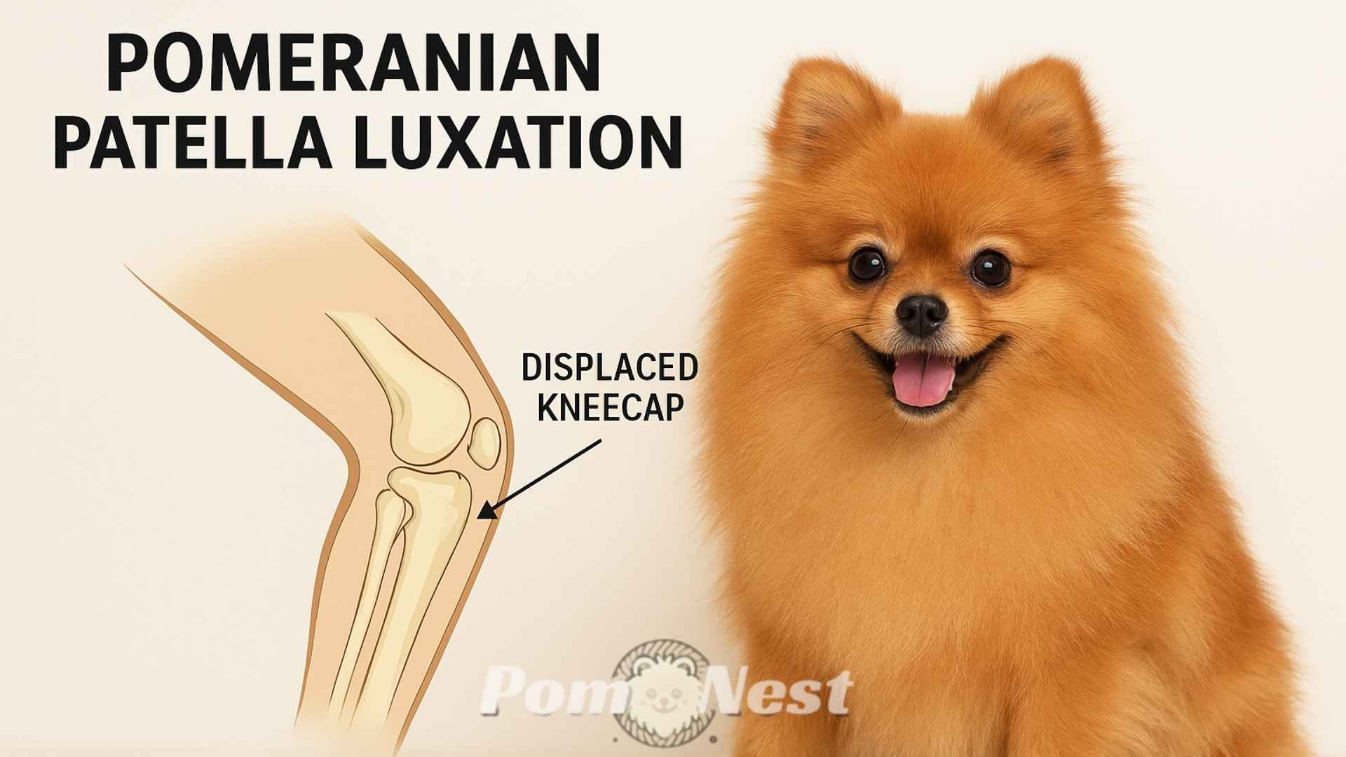
Since LCPD primarily affects small-breed dogs, the veterinarian must rule out other potential causes of rear-leg lameness, such as patellar luxation (slipping kneecap) or soft-tissue injuries. The X-rays are the best tool for this, as the unique bone destruction of Legg Calve Perthes Disease in Dogs is unmistakable.
Treatment of Legg Calve Perthes Disease in Dogs

The treatment approach depends on the severity of the joint damage and how long the dog has had the condition.
Conservative vs. Surgical Options
In very mild, early cases, your vet may recommend a conservative approach, including strict rest and the use of pain relief medication, such as Non-steroidal anti-inflammatory drugs (NSAIDs) for dogs. However, because LCPD usually causes severe damage quickly, surgery is generally the most effective and recommended course of action for long-term comfort.
Femoral Head Ostectomy (FHO) Explained
The most common surgical solution for LCPD is the Femoral head and neck ostectomy (FHO). During this procedure, the surgeon removes the damaged femoral head and neck, effectively eliminating the painful bone-on-bone contact. The resulting void heals to form a “false joint” made of scar tissue, allowing the dog to regain pain-free mobility.
The FHO Procedure Step-by-Step
The FHO procedure involves a surgical incision over the hip, exposing the joint capsule.
The surgeon then carefully cuts away the entire femoral head and neck.
After removing the damaged bone, the muscles and soft tissues are sutured, and rehabilitation begins immediately to encourage the formation of a robust false joint.
Total Hip Replacement (THR) as an Alternative
In certain cases, particularly in slightly larger or more athletic small dogs, Total hip replacement in dogs (THR) may be an option. This procedure replaces the damaged ball and socket with prosthetic implants. While THR offers superior joint function, it is significantly more expensive, more complex, and typically only performed by specialized veterinary surgeons.
What happens to the joint if part of the hip is removed?
When the femoral head is removed via FHO, the body forms a cushion of scar tissue and muscle between the remaining thigh bone and the pelvis. This acts as a fibrous pseudo-joint. The dog moves by relying on the surrounding muscles to support the limb. While the leg may not have perfect range of motion, the procedure successfully removes the primary source of excruciating pain.
Can complications occur with the surgery?
Yes, complications can occur, although they are rare and usually minor. Potential complications of FHO include infection, nerve damage, or, most commonly, insufficient rehabilitation leading to stiffness. It’s imperative that you follow the post-operative instructions precisely to ensure your dog achieves the best functional outcome.
Recovery and Management of Legg Calve Perthes Disease in Dogs

Recovery is just as important as the surgery itself. A committed owner plays the biggest role in ensuring a complete and functional recovery for the dog after FHO.
Immediate Post-Operative Care and Pain Management
For the first few days after the FHO or THR, your dog will require strict rest and careful monitoring. Your vet will prescribe pain medication, including NSAIDs, to manage residual joint pain in dogs and swelling. Gentle passive range-of-motion exercises often begin within a week to prevent scar tissue from becoming too tight.
The Importance of Physical Rehabilitation (Physiotherapy)
Rehabilitation is non-negotiable for FHO success. Canine physical therapy starts early and is crucial for rebuilding the wasted thigh muscle. Exercises include controlled walking, swimming, and specific range-of-motion drills. This intensive work helps the dog properly utilize the new pseudo-joint and prevents permanent canine lameness.
Long-Term Outlook and Quality of Life
The prognosis for dogs treated surgically for LCPD is excellent. Most dogs, especially small breeds, return to a normal, pain-free life and can participate in regular activities. You won’t usually notice any reduced range of motion impacting their day-to-day comfort once they fully recover from Legg Calve Perthes Disease in Dogs.
Will I be able to breed my dog?
Veterinarians strongly discourage breeding any dog diagnosed with LCPD, regardless of the treatment success. Since the condition has strong genetic factors, breeding an affected dog risks passing the painful and debilitating trait to the next generation of puppies.
Prevention of Legg Calve Perthes Disease in Dogs

While you can’t guarantee prevention against a genetic disease, you can take steps to reduce the likelihood and severity of the condition in at-risk breeds.
Responsible Breeding Practices and Screening
The most effective prevention strategy involves responsible breeding. Reputable breeders should actively screen their breeding stock, particularly those popular small-breed dogs known to carry LCPD. Any dog diagnosed with the condition should be immediately removed from the breeding pool to eliminate the hereditary link.
Nutritional Considerations in Puppyhood
Providing your puppy with a high-quality, balanced diet is always recommended for optimal bone health. However, avoiding over-nutrition or excessive supplementation is also important. Rapid, uncontrolled growth can sometimes put undue stress on developing joints. Consulting with your veterinarian about appropriate growth rates for your small-breed dog is a wise proactive measure.
⚠️ Important Veterinary Disclaimer
The content of this article is for informational purposes only and is not intended to be a substitute for professional veterinary advice, diagnosis, or treatment. Always seek the advice of your veterinarian or other qualified animal health provider with any questions you may have regarding a medical condition. Never disregard professional veterinary advice or delay in seeking it because of something you have read here.
References:
- Legg Calve Perthes Disease in Dogs(Royal Canin Academy)
- Legg-Calvé-Perthes Disease (Veterinary Specialty Center of Sarasota)
- Legg Calve Perthes Disease in Dogs (PetMD)
- Understanding Legg Calve Perthes Disease (Dogs NSW)
- Legg Calvé Perthes disease in dogs (PDSA)
- Legg-Calve-Perthes Disease (VCA Animal Hospitals)
- Legg-Perthes Disease in Dogs (Veterinary Partner – VIN)
People also ask:
Perthes disease often requires treatment—either surgical (FHO) or strict conservative management—to heal properly, as the body’s attempt to replace the dead bone can result in a severely deformed, painful femoral head if left unchecked.
Other orthopedic conditions presenting with hindlimb lameness in small-breed puppies can be mistaken for LCPD, most notably patellar luxation (slipped kneecap), septic arthritis, or early signs of hip dysplasia.
LCPD is severely painful, progressing from mild discomfort to non-weight-bearing lameness as the blood-starved bone of the femoral head fragments and collapses, causing bone-on-bone grinding and secondary inflammation.
During the symptomatic phase before treatment, all high-impact activities such as running, jumping, and strenuous play must be strictly avoided to prevent further collapse of the fragile femoral head.
No, LCPD is an aseptic (non-infectious) necrosis and typically does not cause a fever; the presence of a fever would strongly suggest a different condition, like an infectious septic arthritis.
You may also like


Build the Perfect Care Routine for Your Pomeranian

Select your Pom’s age, coat type, and activity level to instantly get a custom care checklist — it’s fast, free, and made with love!
Give your Pom the daily care they truly deserve.